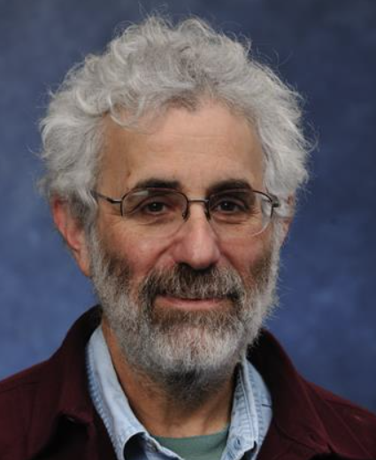
George Stetten, MD, PhD
Mentor

Dr. George Stetten
(Bioengineering, Radiology, CMU Robotics Institute) Visualization techniques for guiding interventional devices; Automated image analysis for feature-based identification and measurement of anatomical structure and motion
Our current research focuses on two main areas. The first involves developing a new device for image-guided intervention called the Sonic Flashlight ™, which provides a stable virtual image of an ultrasound scan within the patient in real time. The device is currently being tested on phantoms and is planned for testing in humans, with the first clinical application the insertion of Peripherally Inserted Central Catheter (PICC) lines into the upper arm. Extensions of this technology to remote and magnified image guided intervention, including light microscopy, as well as to holography, are also being developed. We have recently developed a version of the Sonic Flashlight that uses Real Time 3D ultrasound, an imaging modality Dr. Stetten helped to develop at Duke University in the 1990's. Our second area of interest is in model-based image analysis techniques for automated identification and measurement of anatomical structure and motion, primarily of the heart. We are developing unsupervised model building techniques to accomplish this, using medial-based image features that report the scale and shape of local object centers. We describe more complex structures using the geometric relationships between these medial features. Our group is one of the founding members of the National Library of Medicine (NLM) software consortium creating the Insight Toolkit (ITK, see www.itk.org ), an open-source software library for medical image analysis. We have funding through the NLM for, among other things, the development of ITK as a teaching tool in a new course taught jointly between Bioengineering and the CMU Robotics Institute. In addition to these two main areas, we are exploring new prosthetic devices for the blind, electronic musical instrument design, and other devices in the context of a new practical electronics curriculum in the Bioengineering Department.
